What is it?
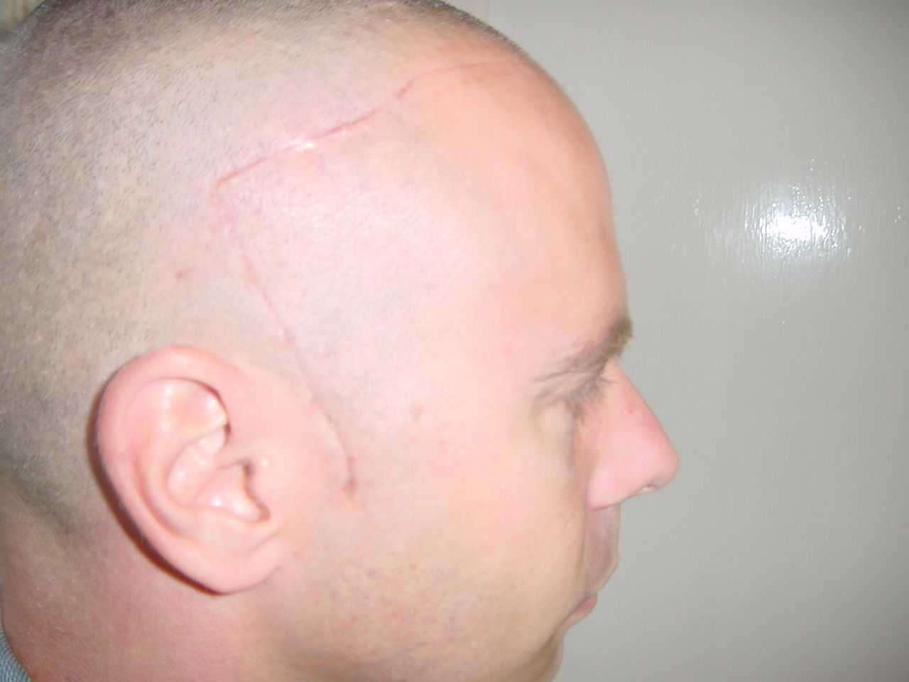
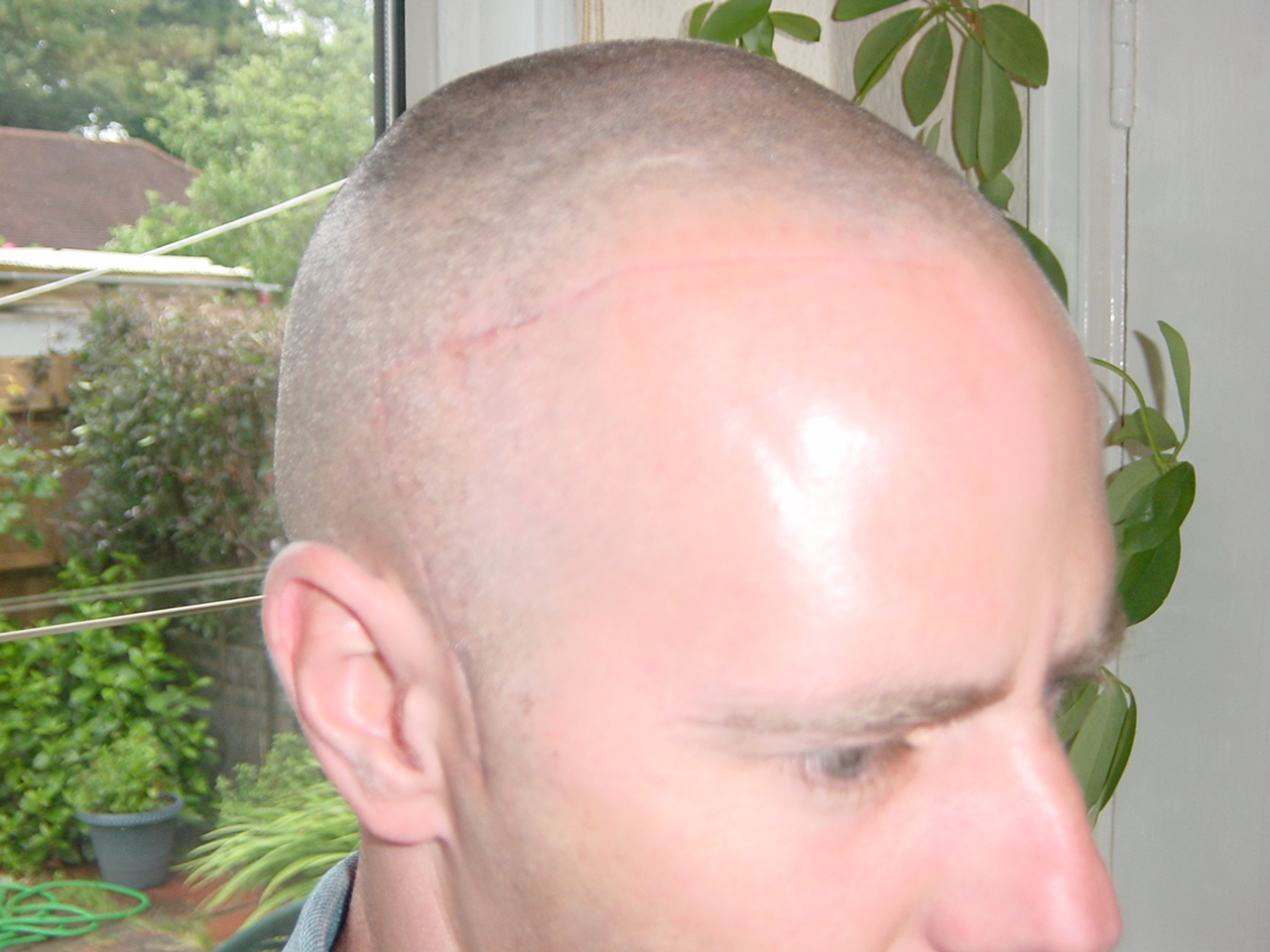
<<<<< May 2004 Operation & Pictures >>>>>
*** Subarachnoid Haemorrhage ***
I was admitted to Ashford Hospital on 15th May 2004 and was diagnosed with a Subarachnoid Haemorrhage. I was then transferred to Charring Cross Hospital for a Craniotomy & clipping of an Aneurysm. The pictures below show the scars I had following the surgery to my brain.
What is it?
A subarachnoid haemorrhage is a serious, potentially life-threatening condition. It happens when an artery close to the brain surface ruptures. Blood leaks out into the space between the membranes that cover the brain and spinal chord.
It is a very rare condition (there are less than 10,000 in the UK every year) although it is always serious and needs urgent medical attention. Most people affected are between the ages of 35 and 65. However, it is not possible to tell who is at risk of a subarachnoid haemorrhage.
There are three membranes that cover the brain and spinal chord. Together they are known as the Meninges. They are: Pia mater (inner membrane), Arachnoid mater (middle membrane), and Dura mater (outer membrane).
Between the inner membrane and the middle membrane is a network of blood vessels which are surrounded by a clear fluid (known as cerebrospinal fluid). In a subarachnoid haemorrhage, one of these blood vessels bursts and blood leaks out into the cerebrospinal fluid.
Diagnosis…..
An initial diagnosis is usually based on the characteristic symptoms, but frequently, other tests are also used. These include:
· Computed tomography scan (CT scan): this can show where the bleeding is and how much blood has leaked between the inner and middle membranes.
· MRI (Magnetic Resonance Imaging) and / or angiogram (a special type of x-ray): this is used to look at the network of blood vessels and can show the berry aneurysm if this is the cause of the haemorrhage.
· Lumbar puncture: this is a test to look at cerebrospinal fluid. If there is a subarachnoid haemorrhage, then there will be traces of blood in the fluid. This is not used very often.
These tests are used to confirm that a subarachnoid haemorrhage has taken place and to make decisions about the most appropriate treatment option.
Symptoms…..
The common symptoms of a subarachnoid haemorrhage are:
· Headache: usually this comes on very quickly and is severe
· Feeling sick and vomiting
· Feeling sleepy or losing consciousness
· Seizures (fits)
· Confusion
· Other symptoms may include dislike of bright light and a stiff neck.
The symptoms can vary between different people will also depend on how serious the subarachnoid haemorrhage is. Sometimes the symptoms can be similar to meningitis (an infection of the Meninges) but both conditions are very serious and medical attention should be sought as soon as possible.
Treatment….
If the subarachnoid haemorrhage has been caused by a berry aneurysm, then brain surgery will probably be needed. The operation involves using a small metal clip to seal off the aneurysm so that it cannot bleed again. The surgery is carried out by shaving the head and opening up the skull to access the brain. This is known as a craniotomy.
Sometimes an alternative treatment is used which involved accessing the berry aneurysm via an artery (using keyhole surgery techniques). A small metal coil is then placed at the centre of the aneurysm. This is known endovascular coiling or embolisation.
A neurosurgeon (brain surgeon) will advise on the most appropriate treatment for a specific patient.
If the subarachnoid haemorrhage has been caused by a malformed artery or vein, then the malformation will be completely removed by a craniotomy.
Subarachnoid haemorrhage is a serious condition and the treatments for it often involved complex brain surgery. For this reason, people with a subarachnoid haemorrhage may be looked after in a hospital intensive care ward (also known as critical care wards)
Any surgery will probably be carried out in a special neurosurgery unit where surgeons have the special skills needed.
Drugs, such as calcium channel blockers, may also be used to keep blood pressure down, reduce the risk of stroke, and to reduce the risk of complications.
<< Back To Top >>
© Keith Humphreys 2004 <<<<< khumphreys2002@yahoo.co.uk >>>>>
|
|